April 2024
 The University Hospital Bonn introduces a transformative mixed-reality
system for thoracic surgery. This innovative approach employs
holographic overlays and real-time 3D imaging to refine surgical
planning for chest wall procedures. By enabling detailed preoperative
visualization of a patient's anatomy, this technology aims to improve
surgical outcomes and enhance patient care.
Arensmeyer, J.; Bedetti, B.; Schnorr, P.; Buermann, J.; Zalepugas, D.;
Schmidt, J.; Feodorovici, P. A System for Mixed-Reality Holographic
Overlays of Real-Time Rendered 3D-Reconstructed Imaging Using a Video
Pass-through Head-Mounted Display—A Pathway to Future Navigation in
Chest Wall Surgery. J. Clin. Med. 2024, 13, 2080.
https://doi.org/10.3390/jcm13072080
Introduction to a Surgical Revolution
In a remarkable stride forward for thoracic surgery, a team of
researchers at the University Hospital Bonn, has developed a pioneering
system that employs mixed-reality and 3D imaging to improve surgical
planning, especially for intricate chest wall surgeries. Their study,
"A System for Mixed-Reality Holographic Overlays of Real-Time
Rendered 3D-Reconstructed Imaging Using a Video Passthrough
Head-Mounted Display" , not only opens new avenues in pre-surgical planning but also sets the
stage for enhanced surgical precision and patient outcomes.
The University Hospital Bonn introduces a transformative mixed-reality
system for thoracic surgery. This innovative approach employs
holographic overlays and real-time 3D imaging to refine surgical
planning for chest wall procedures. By enabling detailed preoperative
visualization of a patient's anatomy, this technology aims to improve
surgical outcomes and enhance patient care.
Arensmeyer, J.; Bedetti, B.; Schnorr, P.; Buermann, J.; Zalepugas, D.;
Schmidt, J.; Feodorovici, P. A System for Mixed-Reality Holographic
Overlays of Real-Time Rendered 3D-Reconstructed Imaging Using a Video
Pass-through Head-Mounted Display—A Pathway to Future Navigation in
Chest Wall Surgery. J. Clin. Med. 2024, 13, 2080.
https://doi.org/10.3390/jcm13072080
Introduction to a Surgical Revolution
In a remarkable stride forward for thoracic surgery, a team of
researchers at the University Hospital Bonn, has developed a pioneering
system that employs mixed-reality and 3D imaging to improve surgical
planning, especially for intricate chest wall surgeries. Their study,
"A System for Mixed-Reality Holographic Overlays of Real-Time
Rendered 3D-Reconstructed Imaging Using a Video Passthrough
Head-Mounted Display" , not only opens new avenues in pre-surgical planning but also sets the
stage for enhanced surgical precision and patient outcomes.
 Innovation at the Core
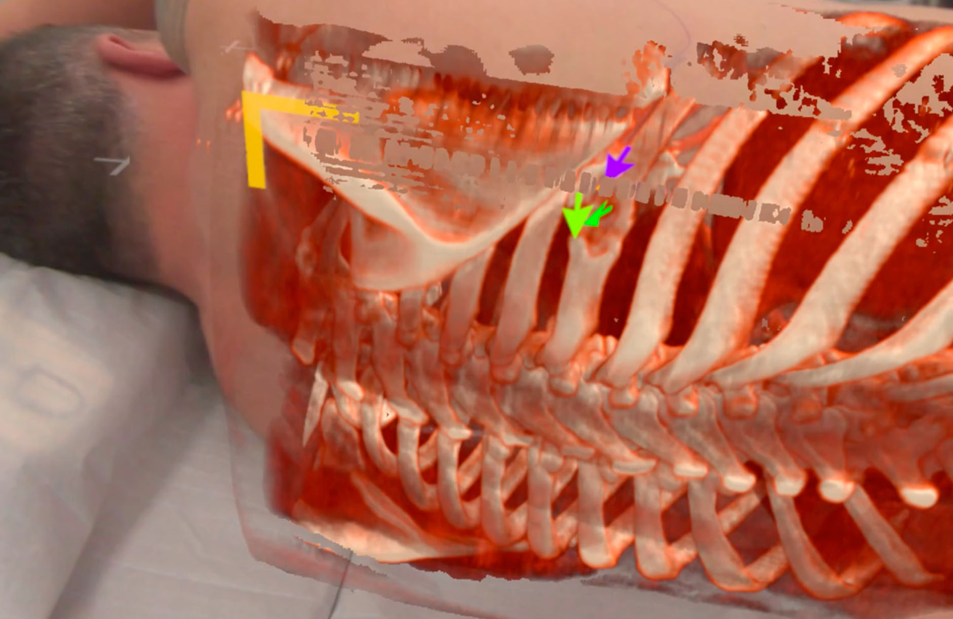
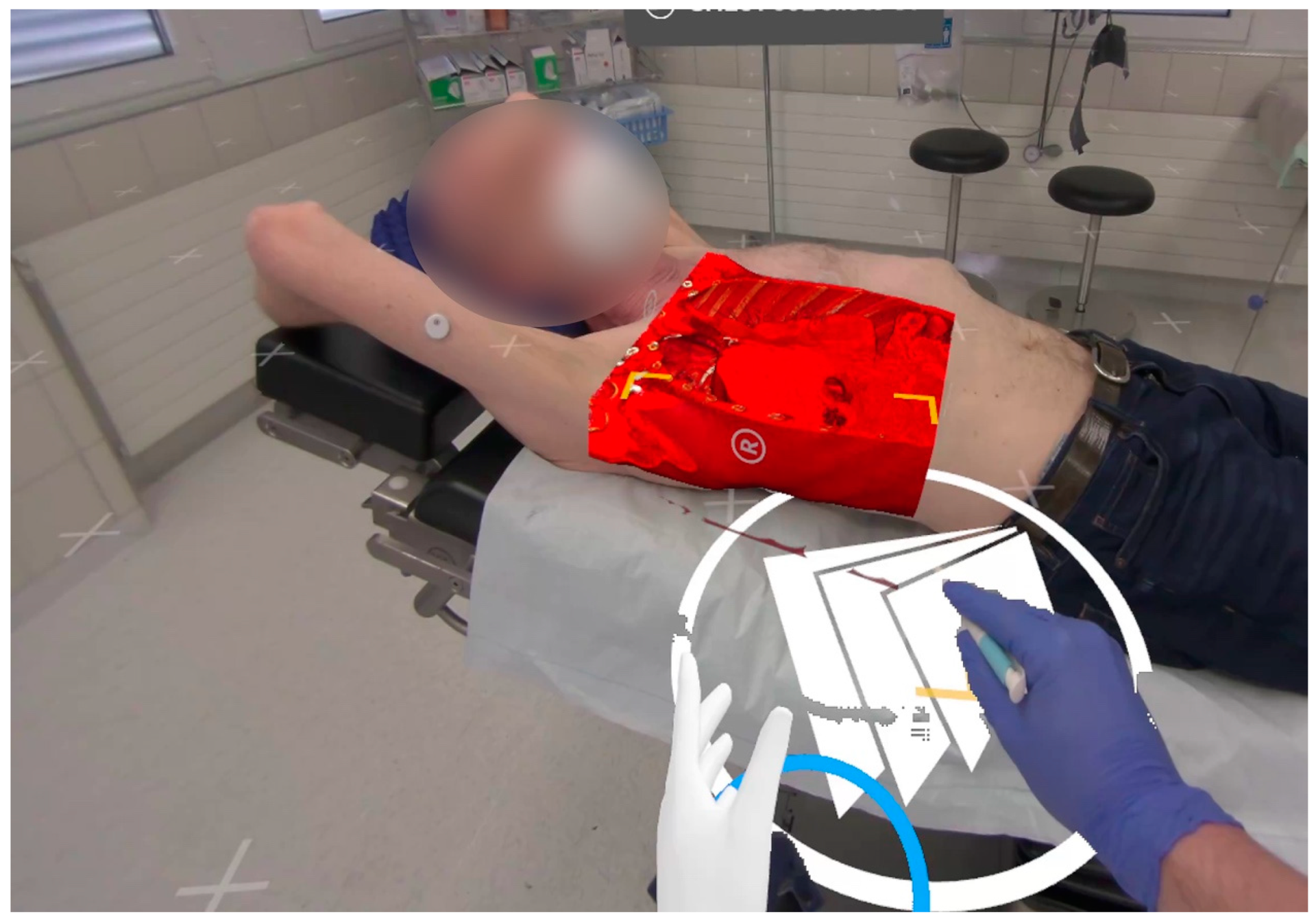
The crux of this innovation lies in the use of high-resolution imaging,
reconstructed in three dimensions, to create a holographic projection of
the patient's anatomy directly onto their body. This allows surgeons to
visualize and interact with the anatomical structures in real-time,
thereby offering a clearer understanding of the surgical site before
making the first incision. The system leverages a state-of-the-art video
pass-through head-mounted display, connected to a high-performance
workstation, to render these images in real-time, enabling surgeons to
manipulate and explore various surgical scenarios and approaches.
The study showcased the efficacy of this system through three
oncological cases, each presenting unique challenges due to the size and
location of the tumors. By projecting the 3D holographic images onto the
patients, surgeons were able to gain invaluable insights into the
spatial relationships between the tumors and critical anatomical
structures, thus facilitating more informed decision-making and surgical
planning.
Holographic Precision in Surgical Planning
This mixed-reality approach marks a significant departure from
traditional preoperative planning methods, which often rely on 2D
imaging or static 3D models. The dynamic nature of the holographic
overlays, combined with the intuitive interaction provided by the
mixed-reality environment, offers a more comprehensive understanding of
the patient's anatomy, which is particularly crucial in complex surgical
tasks within the thoracic cavity.
Innovation at the Core
The crux of this innovation lies in the use of high-resolution imaging,
reconstructed in three dimensions, to create a holographic projection of
the patient's anatomy directly onto their body. This allows surgeons to
visualize and interact with the anatomical structures in real-time,
thereby offering a clearer understanding of the surgical site before
making the first incision. The system leverages a state-of-the-art video
pass-through head-mounted display, connected to a high-performance
workstation, to render these images in real-time, enabling surgeons to
manipulate and explore various surgical scenarios and approaches.
The study showcased the efficacy of this system through three
oncological cases, each presenting unique challenges due to the size and
location of the tumors. By projecting the 3D holographic images onto the
patients, surgeons were able to gain invaluable insights into the
spatial relationships between the tumors and critical anatomical
structures, thus facilitating more informed decision-making and surgical
planning.
Holographic Precision in Surgical Planning
This mixed-reality approach marks a significant departure from
traditional preoperative planning methods, which often rely on 2D
imaging or static 3D models. The dynamic nature of the holographic
overlays, combined with the intuitive interaction provided by the
mixed-reality environment, offers a more comprehensive understanding of
the patient's anatomy, which is particularly crucial in complex surgical
tasks within the thoracic cavity.
 Proven Efficacy in Complex Cases
Moreover, the system's potential extends beyond preoperative planning.
In the future, such technology could be integrated into actual surgical
procedures, offering real-time guidance and navigation, thereby
minimizing the reliance on intraoperative imaging and reducing the
exposure to radiation. This is particularly relevant for smaller lesions
or in minimally invasive and robotic-assisted surgeries, where tactile
feedback is limited.
Challenges Ahead and Future Possibilities
However, the path to widespread adoption of this technology in surgical
settings is not without challenges. The current setup, while advanced,
requires a tethered connection to a workstation, limiting mobility.
Additionally, regulatory hurdles need to be addressed, especially for
intraoperative use. Yet, with rapid advancements in mixed-reality
hardware and cloud computing, these barriers are likely to diminish,
making such sophisticated systems more accessible and versatile for
surgical applications.
The study not only underscores the significant potential of
mixed-reality and real-time 3D imaging in enhancing surgical planning
and execution but also highlights the need for further research to
explore the full scope of its applicability, accuracy, and impact on
clinical outcomes. As we stand on the brink of a new era in surgical
technology, it's clear that the integration of mixed-reality systems
could redefine the standards of surgical precision and patient care in
thoracic surgery and beyond.
For more information, contact
info@medicalholodeck.com
Proven Efficacy in Complex Cases
Moreover, the system's potential extends beyond preoperative planning.
In the future, such technology could be integrated into actual surgical
procedures, offering real-time guidance and navigation, thereby
minimizing the reliance on intraoperative imaging and reducing the
exposure to radiation. This is particularly relevant for smaller lesions
or in minimally invasive and robotic-assisted surgeries, where tactile
feedback is limited.
Challenges Ahead and Future Possibilities
However, the path to widespread adoption of this technology in surgical
settings is not without challenges. The current setup, while advanced,
requires a tethered connection to a workstation, limiting mobility.
Additionally, regulatory hurdles need to be addressed, especially for
intraoperative use. Yet, with rapid advancements in mixed-reality
hardware and cloud computing, these barriers are likely to diminish,
making such sophisticated systems more accessible and versatile for
surgical applications.
The study not only underscores the significant potential of
mixed-reality and real-time 3D imaging in enhancing surgical planning
and execution but also highlights the need for further research to
explore the full scope of its applicability, accuracy, and impact on
clinical outcomes. As we stand on the brink of a new era in surgical
technology, it's clear that the integration of mixed-reality systems
could redefine the standards of surgical precision and patient care in
thoracic surgery and beyond.
For more information, contact
info@medicalholodeck.com