Dissection Master XR is the human anatomy atlas and dissection lab in virtual reality. Based on real human bodies, the software visualizes the human anatomy in a superb level of detail in a fully immersive holographic space. Dissection Master XR is used around the globe for anatomy and medical teaching.
Studying complex human anatomy in virtual reality is easier, faster, and more accurate than with conventional methods on a flat screen or in books. Walk around the dissections, grab the body with your hands, enlarge them to any size, and dive into the smallest anatomical structures in VR.
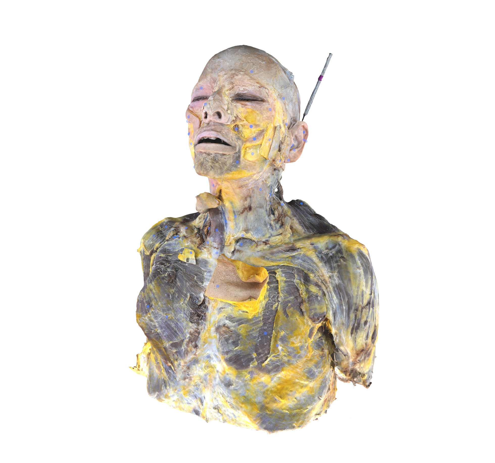
The human body is displayed in ten hi-res layers. You can browse from the outer muscles to the inner organs and study and teach thousands of anatomical structures in utmost detail.
With Dissection Master XR, human anatomy can be visualized, studied, and understood faster and better than ever before. And you can create your own lessons and simulations in VR. Record your teachings for replay in VR, and build a growing library of your anatomy lessons.
Dissection Master XR leads to an improved understanding of human anatomy and physiology and creates a new generation of highly skilled medical professionals.
Start teaching anatomy with Dissection Master XR now!
Experience the human body and its anatomy in a fully immersive holographic space. Welcome to Dissection Master XR, your human anatomy atlas in VR.
The whole human body - from head to toe - is offered in superb quality in ten immersive and fully annotated layers.
More than 3000 anatomical structures are named, annotated, and linked to additional information.
Enlarge the human body, and examine and study it from any angle and perspective. And compare it with medical imaging and Zygote's anatomy data using the other Medicalholodeck apps.
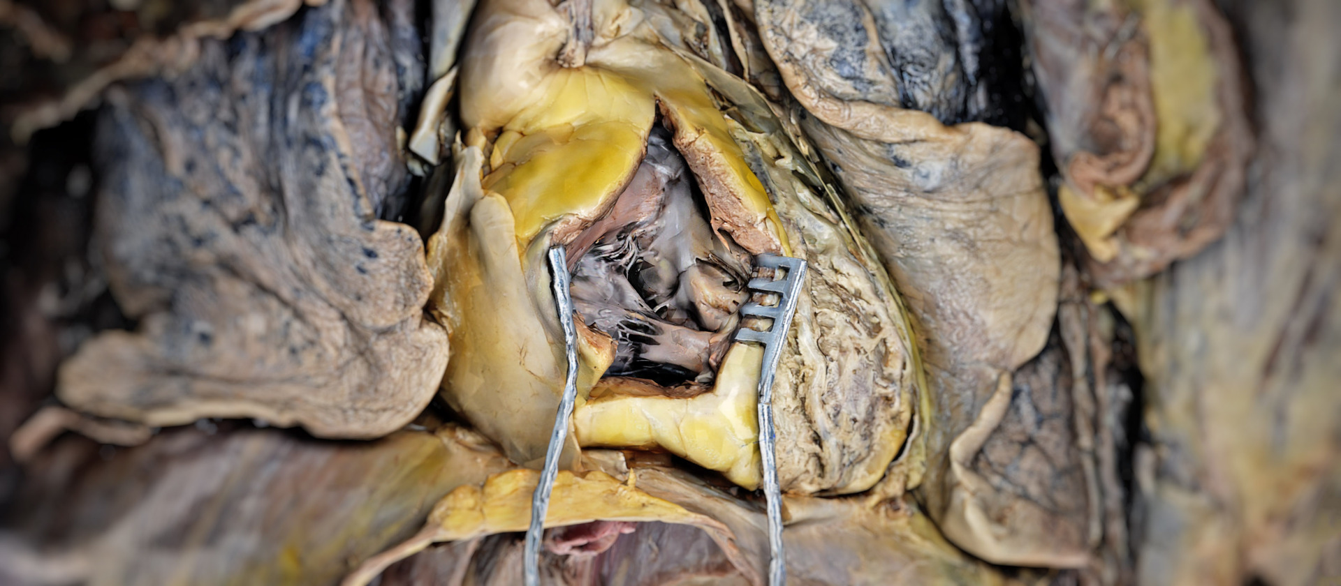
The dissections are carefully created by a leading specialist.
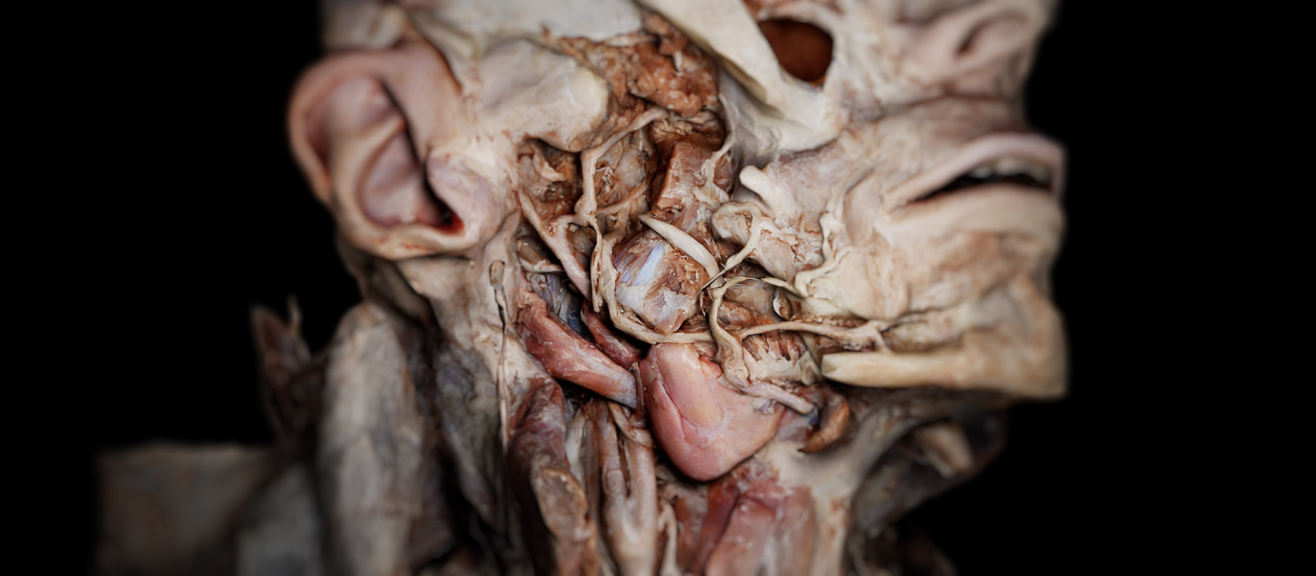
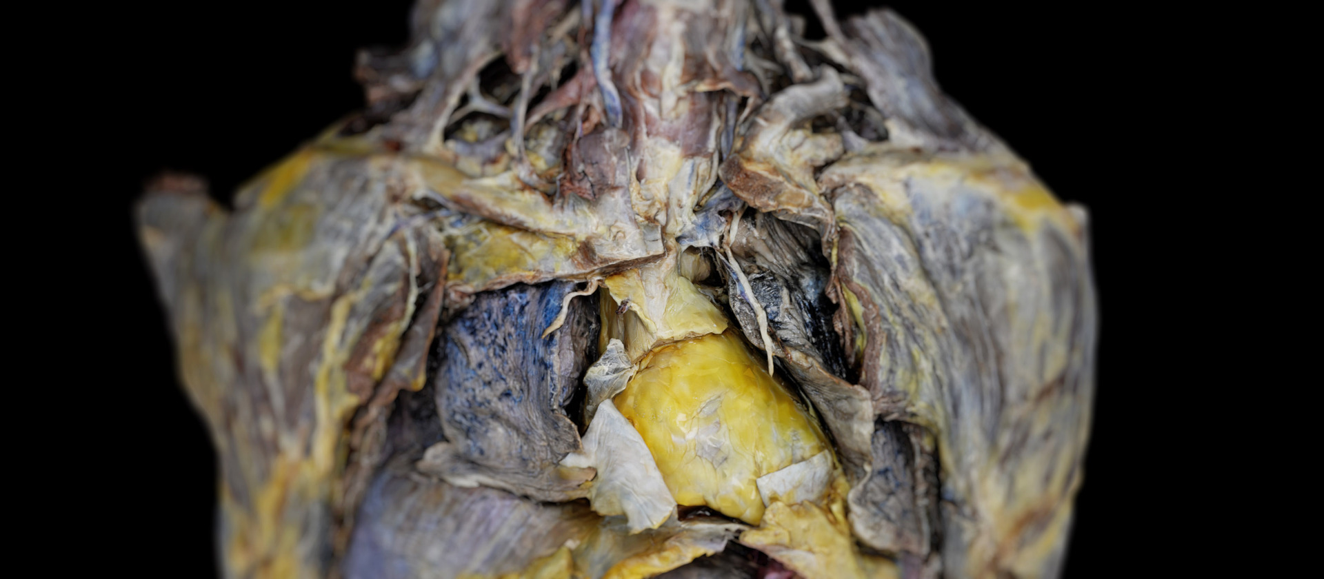
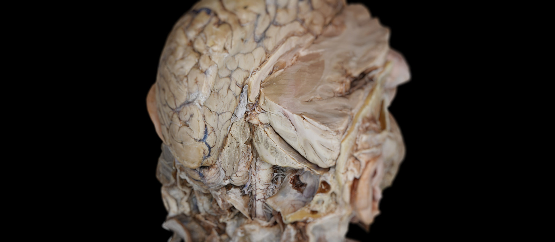
Real human dissections form the basis of the atlas. After dissection, the body was carefully digitized using the latest state-of-the-art technology.
Teach human anatomy at your department and location-independent in virtual classrooms in the metaverse.
Record anatomy lessons, develop your content, and store and share your know-how with users worldwide.
The abdominal aorta, stretching downward from the thoracic aorta, serves as a pivotal vessel supplying oxygen-rich blood to numerous abdominal organs. With indispensable roles in bodily function and circulatory health, it bifurcates at its terminus, forming two iliac arteries, which subsequently ensure the vitality of the lower extremities by providing them with a rich blood supply.
Nestled between thorax and pelvis, the abdominal cavity constitutes a significant component of human anatomy. Within its confines, numerous vital organs, such as the liver, stomach, and intestines, find residence. Surrounded by protective muscular structures and internally lined with the peritoneum, it plays a critical role in various life-sustaining physiological processes.
Dispersed throughout the abdominal region, the abdominal lymph nodes tirelessly guard against infection and disease. These tiny, bean-shaped structures are responsible for filtering lymph fluid, intercepting and disabling potential threats like pathogens. Ensuring effective lymphatic flow, they remain a crucial component of the immune response.
Embedded in both the hand and foot, the abductor digiti minimi muscle contributes to the mobility and functionality of the little finger and toe. In the hand, it enables the pinky finger to abduct, while in the foot, it facilitates the lateral movement of the little toe. It is crucial for maintaining balance and enabling delicate manipulative activities..
Positioned in the foot, the abductor hallucis muscle performs the essential task of enabling big toe abduction. Engaging in moving it away from the other toes, its functionality is most prominent during ambulation and when maintaining equilibrium. Its role, though subtle, significantly impacts overall mobility and stability in locomotion.
Located in the hand, the abductor pollicis brevis is dedicated to facilitating the nuanced movements of the thumb. It abducts the thumb, moving it away from the hand’s palm, which is paramount for various gripping actions and manual manipulations. The utility of this muscle is exemplified in everyday gestures and intricate manual activities.
The abductor pollicis longus, residing further in the forearm, orchestrates precise thumb movements. This muscle not only abducts the thumb but also assists in its rotation. By doing so, it significantly enhances the thumb's dexterity and functionality, which is vital for performing a plethora of manual tasks and expressive gestures.
Situated in the thoracic area, the accessory hemi-azygos and superior hemi-azygos veins engage in the vital task of draining blood from parts of the thoracic wall and upper lumbar regions. These veins ensure the efficient return of deoxygenated blood back to the heart, playing a subtle yet crucial role in the circulatory system.
Belonging to the illustrious family of cranial nerves, the Accessory Nerve, also termed Cranial Nerve XI, plays a significant role in the human body. Comprising both cranial and spinal segments, this nerve is primarily involved with controlling muscles in the neck and shoulder, like the sternocleidomastoid and trapezius. Its function is keenly felt when turning the head or shrugging the shoulders, illustrating its importance in daily movements.
An interesting feature of human anatomy is its variability, exemplified by the Accessory Obturator Artery. Not universally present, when it does make an appearance, it often originates from the inferior epigastric artery. This artery, though a variant, remains important by supplying blood to areas around the hip and pelvic region, reflecting the human body's adaptability.
Complementing the Accessory Obturator Artery, the Accessory Obturator Vein is responsible for draining blood from regions supplied by the aforementioned artery. This ensures that the pelvic region enjoys efficient venous return, keeping the area well-circulated and in optimal health.
A variation of the widely recognized great saphenous vein, the Accessory Saphenous Vein, navigates the terrain of the leg. Despite being a variant, its function remains vital. It focuses on effectively channeling blood from the leg back to the heart, contributing significantly to the leg's venous circulation system.
Encircling the acetabulum of the hip is the Acetabular Labrum, a fibrocartilaginous structure. By amplifying the depth of the acetabulum, it acts as a cushion and boosts the joint's stability. Every step and hip movement benefits from this structure, enhancing the joint's security and facilitating smooth mobility.
An integral component of the hip structure, the Acetabulum is a concave surface on the pelvis. It welcomes the head of the femur, together forming the iconic ball-and-socket hip joint. As a cornerstone of the body's locomotion system, the Acetabulum ensures a wide range of movements, from walking to dancing.
A distinct bony projection from the scapula, the Acromion, connects with the clavicle to give rise to the acromioclavicular joint. Acting as an anchor point for various ligaments and muscles, the Acromion stands as a sentinel ensuring the shoulder's smooth and coordinated movement.
Delving into the inner thigh, one encounters the Adductor Brevis muscle. Specializing in drawing the thigh closer to the body's midline, it's a pivotal part of the adductor muscle group. Whether during a leisurely walk in the park or an intense soccer match, its role in propelling movement is undeniable.
Deep within the foot lies the Adductor Hallucis muscle, dedicated to moving the big toe towards its fellow toes. Its functions aren't just limited to toe movement; it's a key player in maintaining balance and plays a part in the weight distribution dynamics during activities like standing or striding.
Carved within the adductor magnus muscle, the Adductor Hiatus serves as a pivotal passageway. Through this hiatus, the femoral artery and vein travel, transitioning from the thigh region to the popliteal fossa located behind the knee. This opening is a testament to the body's intricate design, ensuring smooth blood circulation.
Situated within the inner thigh, the Adductor Longus is a powerful and elongated muscle. As its name suggests, its primary function is to adduct or draw the thigh inward towards the body's centerline. Integral to actions like crossing legs or making controlled sideways movements, its strategic placement and function underscore its role in daily mobility and stability.
Larger and broader than its counterpart, the Adductor Longus, the Adductor Magnus muscle dominates the medial aspect of the thigh. Its substantial size offers a hint about its purpose, playing a multifaceted role in adduction, flexion, and medial rotation of the thigh. Whether sprinting or simply standing, this muscle's significance is felt across a spectrum of activities.
Understanding human anatomy requires excellent three-dimensional visualizations. Dissection Master XR is your interactive anatomy atlas in virtual reality, offering the world's highest resolution and most detailed anatomical dissections in a fully immersive, holographic space.
Teach and study human anatomy in VR, improve your education's quality, and enhance your students' learning skills.
Connect with your students and teach anatomy in virtual classrooms. Location-independent teaching opens new possibilities for university education, international collaboration, and global learning.
Save time and increase your success. Learning 3D structures in an immersive 3D environment is easier than from flat screens or books.
Create your anatomy lessons, simulations, and content in VR, and make it accessible to users worldwide.
Use Dissection Master XR and Medicalholodeck's data and apps to teach human anatomy in virtual classrooms. Add your media, and create lessons in virtual reality. Build your library of educational content and simulations, and share it with students and users worldwide in the metaverse.
Teach anatomy in VR, add your DICOM, 3D, and 2D files, and create teachings, lessons, and simulations for replay in VR. Use Medicalholodeck to enhance your existing dissection lab and bring your simulation lab to the next level.
Study human anatomy in virtual reality with Dissection Master XR, Anatomy Master XR, and Medical Imaging XR. All applications can be used simultaneously, giving students extraordinary insight into human dissections, anatomy models, and medical imaging. Users confirm that studying in virtual reality saved time and made them more successful in their medical careers.